Histology
Histology
The MIRC histology Core Facility (or John Mayberry Histology Facility) is a fully operational high quality histology core with the capacity to process, embed and section both formalin-fixed and cryopreserved tissues, and stain by chemical, antibody and RNA probe technology. The facility provides support to the members of the McMaster Immunology Research Centre and is open to McMaster Investigators and external laboratories, all on a fee-for service basis.
We offer full service options including:
- processing of formaldehyde-fixed and cryopreserved tissues
- embed, section and stain with standard histological stains
- well developed repertoire of immunohistochemical (IHC) stains
- newly developed capability for mRNA In situ hybridization (ISH)
- slide scanning (light microscopy)
The facility occupies 1000 sq. ft. within the McMaster Immunology Research Centre, equipped with an Tissue-Tek VIP 6A1 Tissue Processor, an Autostainer XL, a CV5030 Automated Coverslipper and transfer station, an EG1150 Embedding Center, two RM2225 microtomes, a Jung Biocut 2035 Microtome and a CM3050 Cryostat. Recent additions to the instrumentation include a Leica Aperio Scanscope XT, and the Leica Bond Rx automated tissue stainer, which provides:
- high quality, reproducible, large capacity IHC.
- RNA Scope ISH technology using chromogenic (CISH) or florescent (FISH) detection
This facility is operated by two highly-experienced certified histologists (Mary Jo Smith and Mary Bruni) with over a decade of running and overseeing the core. Our staff can also work with you to develop new stains. Certain equipment maybe made available for rental on a pay-as-you-go basis.
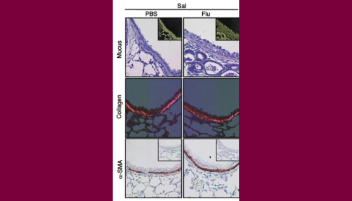
Examples of collagen and mucus staining in addition to smooth muscle cell deposition from: Influenza A facilitates sensitization to house dust mite in infant mice leading to an asthma phenotype in adulthood.
by A Al-Garawi, M Jordana et al.
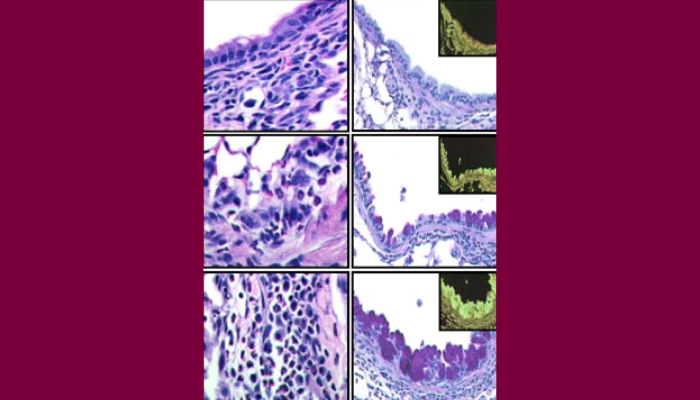
Examples of H&E and periodic acid-Schiff staining to measure lung histopathology after house dust mite exposure in the context of a prior flu infection.
J Immunol. 2009 Mar 1;182(5):3095-104.Al-Garawi et al.
Contact the Histology Facility
For enquiries from outside McMaster, please initially contact the MIRC director, Dr Carl D Richards.

Mary Jo Smith
Head Technician, Histology Facility
McMaster Immunology Research Centre